Foot X-Ray Analysis - Hallux Rigidus
Foot X-Ray Analysis - Hallux Rigidus
Date: March 15, 2026
Analysis by: Claude (AI Medical Image Analysis)
X-Ray Views
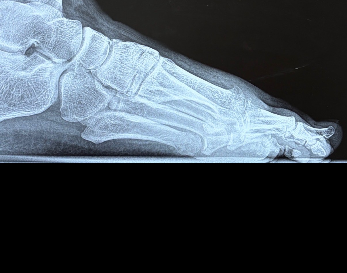
Lateral View
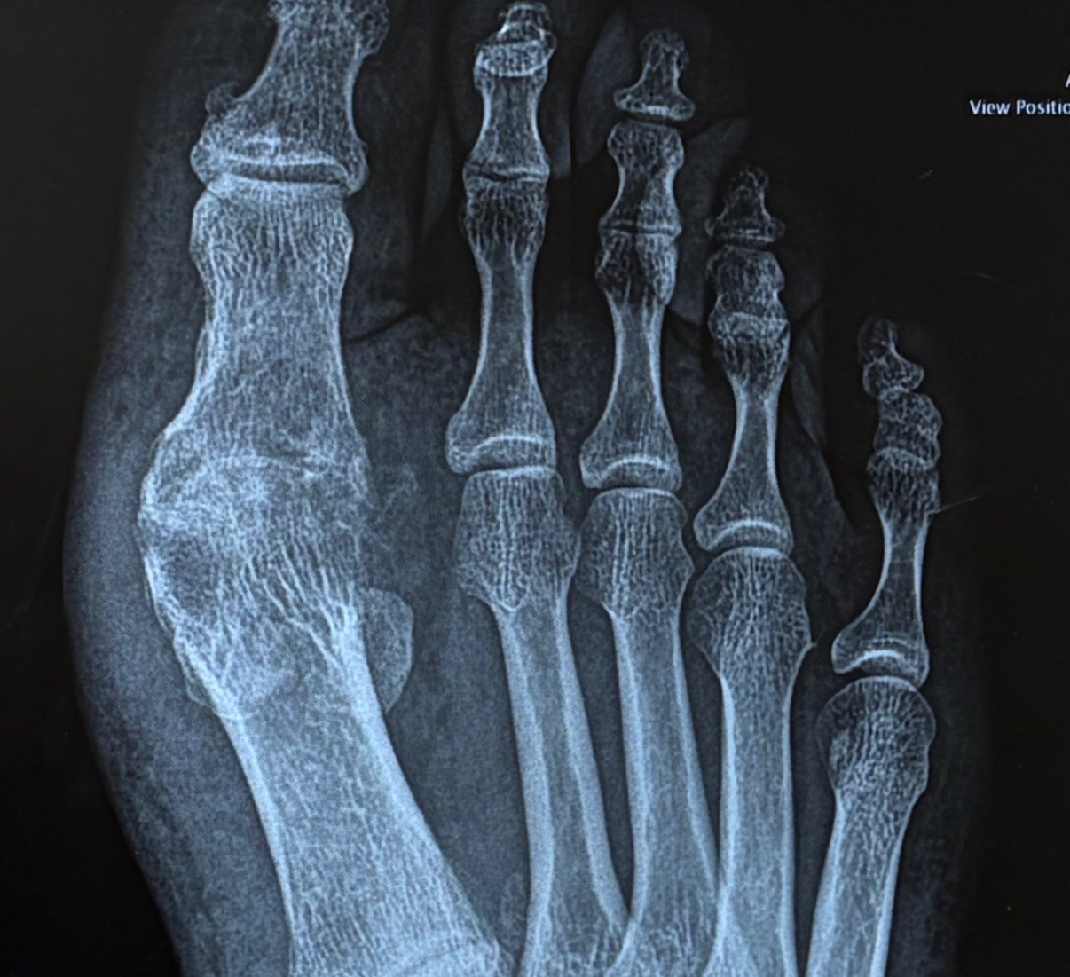
AP (Anteroposterior) View
Clinical Findings
Based on the radiographic images provided, this appears to be a case of hallux rigidus (degenerative arthritis of the first metatarsophalangeal joint).
Key Observations:
1. Joint Space Narrowing - Significant narrowing of the first MTP (metatarsophalangeal) joint space - Indicates cartilage loss and degenerative changes - Most prominent at the dorsal (top) aspect of the joint
2. Osteophyte Formation - Visible bone spurs (osteophytes) at the joint margins - Particularly prominent on the dorsal aspect of the first metatarsal head - Classic sign of osteoarthritis in this location
3. Subchondral Sclerosis - Increased bone density (sclerosis) adjacent to the joint surface - Indicates chronic stress and remodeling - Visible as brighter/whiter bone on the x-ray
4. Joint Alignment - Overall alignment appears maintained - No significant hallux valgus (bunion) deformity - Sesamoid bones appear intact
Hallux Rigidus Overview
Hallux rigidus is a degenerative condition affecting the big toe joint, characterized by:
- Stiffness - Progressive loss of dorsiflexion (upward bending)
- Pain - Especially with walking and push-off
- Bone spurs - Leading to mechanical blocking of motion
- Joint degeneration - Cartilage wear over time
Common Causes:
- Repetitive stress/overuse
- Previous trauma to the joint
- Biomechanical factors (foot structure)
- Genetic predisposition
- Age-related wear and tear
Staging:
Based on radiographic findings, this appears to be Grade 2-3 hallux rigidus: - Grade 1: Mild osteophytes, minimal joint space narrowing - Grade 2: Moderate osteophytes, joint space narrowing, subchondral sclerosis - Grade 3: Severe osteophytes, marked joint space narrowing, cyst formation - Grade 4: Complete joint destruction
Treatment Considerations
Conservative (Non-Surgical): - Stiff-soled shoes or rocker-bottom shoes - NSAIDs for pain/inflammation - Activity modification - Physical therapy - Corticosteroid injections - Orthotic devices with Morton's extension
Surgical Options: - Cheilectomy - Removal of bone spurs (early-stage) - Joint fusion (arthrodesis) - Eliminates motion but relieves pain - Joint replacement - Restores motion (newer option) - Osteotomy - Realignment procedures
Disclaimer
This analysis is for educational purposes only and should not be considered medical advice.
This is an AI-generated interpretation of radiographic images. All medical imaging should be evaluated by qualified healthcare professionals (radiologists, orthopedic surgeons) in the context of clinical examination and patient history. Always consult with your physician or podiatrist for proper diagnosis and treatment recommendations.
Analysis generated by Claude (Anthropic) - March 15, 2026